 |
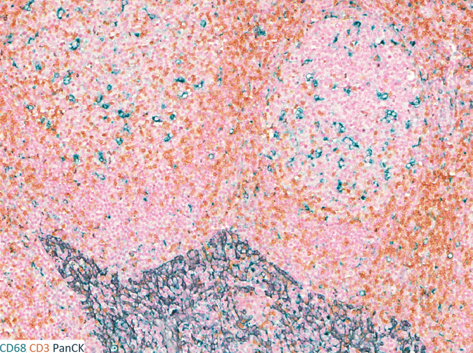 |
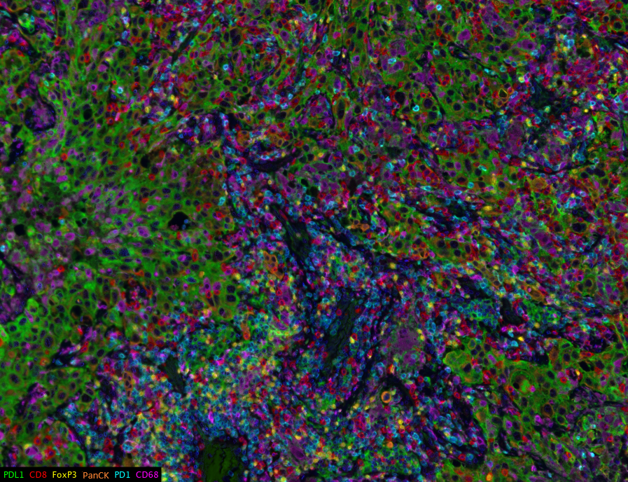
The Tumor Microenvironment (TME) Core performs multiplexed immunofluorescence/immunohistochemical (mIF/mIHC), single color IHC assays, and amplified in situ hybridization (ISH) studies using formalin-fixed paraffin-embedded (FFPE) tissue samples collected from both humans and animals. Additionally, the TME Core will also develop new antibody protocols and mIF/mIHC panels as requested. The TME Core has two Leica Biosystems Bond RX Automated Slide Stainers, and four Akoya Biosciences PhenoImager HT Automated Quantitative Pathology Imaging Systems. The TME also provides access to a Hamamatsu NanoZoomer brightfield slide scanner and a Leica Microsystems LMD7 laser capture microdissection microscope. The software required for image analysis for these platforms (Indicia Labs HALO, inForm Akoya Biosciences, etc) is also be available through the Center.
Please navigate to the request services tab and inititate a request for services through the TME. If you have additional questions please email TMECore@jh.edu
| Janis M. Taube MD |
Robert A. Anders MD, PhD |
| Co-Director | Co-Director |
| Hours | Location |
|
Open 24/7 Staffed M-F 10:00am-4:00pm |
CRB2-216 (Middle Door) 1551 Jefferson Street Baltimore, MD 21287 |
| Name | Role | Phone | Location | |
|---|---|---|---|---|
| Logan Engle |
Sr. Laboratory Manager
|
2-2197
|
TMECore@jh.edu
|
CRB2-216
|
| Service list |
| ► Training (1) | |||
| Name | Description | Price | |
|---|---|---|---|
| LCM Training | Inquire | ||
